
US Images of Acute Appendicitis
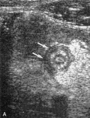
Image 1
Major ultrasonographic findings in acute appendicitis in the RIF include the following:
Major ultrasonographic findings in acute appendicitis in the RIF include the following:
- An aperistaltic, noncompressible, blind-ended, sausage-shaped structure that arises from the base of the cecum
- Distinct appendiceal wall layers
- An outer diameter greater than 6 mm
- A target appearance
- Appendicolith(s) (See the images below.)
- Periappendiceal fluid collection
- Echogenic, prominent pericecal fat
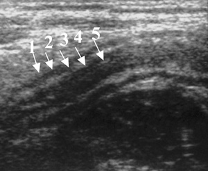
Image 2
The longitudinal ultrasonographic view demonstrates a nonperforated, inflamed appendix that is characterized by an aperistaltic, noncompressible, blind-ended, tubular structure with a laminated wall that arises from the base of the cecum. When the inflammation is mild and visualization is optimal, 5 distinct appendiceal wall layers can be identified (see the image)
